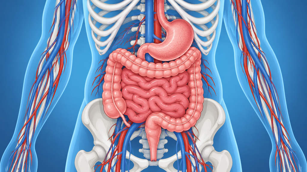
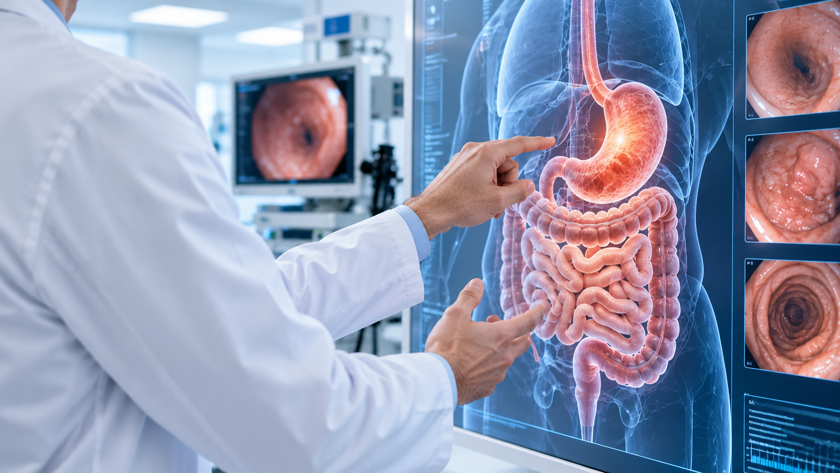
One digestive test can show a seemingly normal result and, even so, not completely resolve the root of a problem. In certain cases, the advanced endoscopy allows areas or abnormalities that may be difficult to identify through a conventional examination to be observed in greater detail.
This does not mean that a gastroscopy, a colonoscopy or an imaging test have been poorly performed or are insufficient. Each test provides different information. Therefore, when symptoms persist, there is a finding that raises doubts or a lesion needs to be studied in greater detail, it may be necessary to resort to techniques of advanced endoscopy that allow a better understanding of what is happening.
What differentiates an advanced endoscopy from a conventional one?
A conventional endoscopy allows direct visualisation of the inside of the digestive tract and the detection of numerous abnormalities in the oesophagus, stomach, duodenum or colon.
The Advanced digestive endoscopy incorporates technologies and procedures that go a step further, principally because it is possible to expand the information on a lesion, study deeper layers of the digestive wall, access certain areas or even obtain tissue samples that are not directly visible from the surface.
Among these techniques are chromoendoscopy, the Ecoendoscopia, the capsule endoscope or certain endoscopic resection techniques, among others.
Its indication always depends on the symptoms, the results of previous tests and the clinical suspicion.
Very small or barely visible mucosal alterations
Some digestive lesions do not form a clearly recognisable polyp or mass. They may be plans, present minimal colour changes or modify the mucosal pattern very subtly.
The use of systems of magnification y image enhancement techniques can facilitate the identification and characterisation of these areas.
This can prove quite useful for studying:
Flat or slightly raised lesions.
Early changes in the digestive mucosa.
Suspicious areas requiring a targeted biopsy.
Abnormalities associated with certain conditions requiring endoscopic surveillance.
It is not solely a matter of “seeing more”, but of better analysing the characteristics of what is being observed.
Early changes in Barrett's oesophagus
The oesophagus Barrettrequires proper follow-up because, in some patients, precancerous cellular changes known as may appear dysplasia.
These alterations do not always form an obvious lesion. Some may manifest as small surface irregularities that require thorough inspection and, when indicated, biopsies to be taken.
The enhanced imaging techniques they help the specialist to locate suspicious areas and direct the study of the mucosa with greater precision.
Correctly identifying these lesions is important because certain early abnormalities may require surveillance, specific biopsies or advanced endoscopic treatments in selected patients.
Lesions beneath the mucosa
An endoscopy can detect a bulge in the stomach or oesophagus, but the superficial image does not always make it possible to determine what lies beneath. These lesions may correspond to subepithelial or submucosal lesions.
In these cases, the Ecoendoscopia it is particularly useful. This technique combines endoscopy with an ultrasound scan performed from inside the digestive tract, making it possible to study the various layers of its wall.
Thanks to it, the following can be analysed:
From which layer does a subepithelial lesion appear to originate.
Its size and characteristics.
His/Her/Their relationship with nearby structures.
If there are elements that advise expanding the study.
If it is necessary to obtain a tissue sample.
In this way, a lesion that from the outside might simply look like a “lump” can be characterised with much greater precision.
Alterations in organs adjacent to the tractor digestive
One of the particularities of the Ecoendoscopia It is not limited to studying the wall of the digestive tract. By performing the ultrasound from the oesophagus, stomach or duodenum, it allows certain nearby structures to be observed at very close range.
For this reason, it can be used, in selected situations, to study lesions or alterations related to the pancreas, the bile duct, certain lymph nodes or other structures close to the digestive system.
In addition, when indicated, it allows samples to be obtained via image-guided puncture, which can provide vital information to reach a diagnosis without initially resorting to more invasive procedures.
Localized problems in the small intestine
The small intestine It is a particularly complex area to study using conventional gastroscopy or colonoscopy, as a large part of its length lies beyond the reach of these examinations.
In certain patients, techniques such as the capsule endoscope, which makes it possible to obtain images of the intestine as a small camera capsule travels naturally through the digestive tract.
It can be useful, for example, for studying certain cases of:
Gastrointestinal bleeding of obscure origin.
Iron deficiency anaemia with no clear cause after initial investigation.
Suspicion of certain diseases of the small intestine.
Injuries requiring a more thorough assessment.
If an abnormality is identified, other endoscopic techniques may be necessary to investigate it, obtain biopsies or carry out treatment.
When is it necessary to extend the study?
Not everyone with digestive symptoms needs one advanced endoscopy. The choice of a more specialised test must respond to a specific indication.
It can arise when:
Symptoms continue despite the initial tests showing no clear cause.
A lesion has been found and it needs to be further characterised.
There is a sub-mucosal lesion.
A suspicious area needs to be studied in detail.
It is necessary to assess whether a lesion can be treated by endoscopy.
It is necessary to obtain tissue from structures that are difficult to reach using other techniques.
Therefore, rather than requesting a specific test in isolation, the important thing is to carry out a specialist valuation that allows the decision of which investigation can genuinely provide useful information. The choice of a more advanced technique depends on the symptoms, previous findings and the clinical suspicion for each patient.
In addition to the technology used, the endoscopist's experience plays a fundamental role. The identification of subtle lesions, the interpretation of certain patterns and the correct characterisation of findings also depend on the training and experience of the specialist. Therefore, Just as important as having advanced techniques is knowing when to use them and how to interpret the information they provide.
Final conclusion: the importance of choosing the right test
A conventional test may be sufficient to diagnose most digestive problems, but there are situations where it is necessary to go deeper. The advanced endoscopy it allows certain lesions to be studied in greater detail, assessing areas that are not accessible with a standard examination and obtaining information on structures located beyond the surface of the digestive tract.
And GASTEIZ we study each case individually to determine which tests are really necessary and when a technique of advanced endoscopy for a more accurate diagnosis. If you have recurring discomfort, an injury that requires a more thorough examination or need a second specialist opinion, you can request a consultation with our Gastroenterology team.
Tell us what you thought of this article. Rate it (FROM 1 TO 5 STARS).