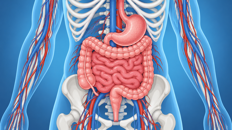




The gastrointestinal submucosal lesions, also commonly known as gastrointestinal submucosal lesions, are findings that may appear during a gastroscopy, a colonoscopy or an imaging test and that, on many occasions, are discovered incidentally. Although many do not produce symptoms and some are completely benign, their external appearance does not always make it possible to know with exactitude what type of lesion it is.
That is why identifying its origin, size and characteristics is essential to decide whether monitoring it is sufficient or if it is advisable to study or treat it.

What are gastrointestinal submucosal lesions?
It is usually called submucosal lesion to a protrusion observed beneath the internal surface of the digestive tract while the mucosa covering it may appear normal. However, from a medical point of view, the term subepithelial lesions it is more precise, since these alterations may originate in different layers of the gastrointestinal wall or even correspond to compression from a structure located outside the digestive tract.
They can be located in the oesophagus, stomach, duodenum, intestine or colon. Their nature is also very varied: some contain fat or muscle tissue, others are cystic and some correspond to tumours with differing growth potential.
Detecting a lesion, therefore, does not automatically equate to diagnosing a specific disease.
What types of injuries can be found?
Inside the gastrointestinal subepithelial lesions very different entities exist. Among them are the lipomas, made up primarily of fatty tissue; leiomyomas, originating in smooth muscle; the ectopic or heterotopic pancreas; certain cysts; the Neuroendocrine tumours and the gastrointestinal stromal tumours or GISTs.
The appearance during endoscopy can guide the specialist, but different lesions can present a similar look. Hence, it is not advisable to draw conclusions solely from their shape or size.
Do they cause symptoms?
Many injuries submucosae digestive they are asymptomatic and are discovered incidentally when performing a endoscopy for another reason. This type of incidental finding is relatively common in endoscopic practice.
However, depending on their location and size, some may cause symptoms. These can include difficulty swallowing if the lesion is located in the oesophagus, abdominal discomfort, a feeling of fullness, digestive bleeding o symptoms related to a partial obstruction.
The presence of ulceration, bleeding or symptoms attributable to the lesion is one of the factors that the specialist will take into account when assessing its management.
Why is making an accurate diagnosis so important?
Two lesions can look very similar during a gastroscopy and yet be completely different. Knowing what type of lesion it really is helps to decide whether it needs treatment, follow-up or simply monitoring.
The study must answer several questions: exactly where the lesion is located, which layer of the digestive wall it originates from, how large it is, whether it is solid or cystic, and whether it has features that make obtaining a tissue sample advisable.
Endosonography, a key test
The Ecoendoscopia It is a technique that combines endoscopy with the use of ultrasound. As a result, it allows the different layers of the digestive wall to be observed in greater detail and provides a better understanding of the characteristics of the lesion.
It is especially useful for knowing the real size of the lesion, determining its source layer, evaluate the margins and analyse their internal structure. Therefore, clinical guidelines consider endoscopic ultrasound to be one of the fundamental tools for characterising subepithelial lesions.
However, endoscopic ultrasound does not always make it possible to establish a definitive diagnosis on its own. In certain cases, it is necessary to obtain tissue for analysis.
When might a biopsy be necessary?
The need to carry out a biopsy It depends on the characteristics of each lesion. When dealing with a solid lesion whose diagnosis is unclear or where there are features requiring histological definition, endoscopic ultrasound-guided tissue acquisition can increase diagnostic accuracy.
One of the available techniques is fine needle biopsy or FNB, which allows small fragments of tissue to be obtained to determine the type of lesion in question. Current recommendations especially consider tissue acquisition when it is necessary to differentiate solid lesions or when there are characteristics that may modify the therapeutic strategy.
Not all lesions require a biopsy. In those with a very characteristic appearance and a clear benign diagnosis, the specialist may consider that further investigation is not necessary.
What treatment do these injuries need?
No. The treatment depends on the diagnosis and the individual risk of each case.
To decide between surveillance, endoscopic resection or surgical treatment, factors such as the following are evaluated:
- Type of lesion and histological diagnosis.
- Size and location.
- Layer of the digestive wall in which it originates.
- Presence of symptoms, ulceration or bleeding.
- Changes observed during follow-up.
- Features associated with a higher malignant potential.
Some correctly identified benign lesions may not require follow-up. Others require regular monitoring via endoscopy or echoendoscopy, whilst certain lesions may benefit from resection.
In the case of the GIST, for example, size, location and other characteristics condition the strategy. Clinical guidelines recommend assessing the resection of gastric GISTs larger than 2 cm and GISTs located outside the stomach, whereas the management of smaller gastric lesions should be individualised.

Conclusion: The key lies in correctly identifying the lesion
The gastrointestinal submucosal lesions can have very different characteristics and evolutions. That is why identifying them correctly is essential to know if they need follow-up, a biopsy or some kind of treatment. Tests such as the Ecoendoscopia they allow them to be studied in greater detail and to make decisions tailored to each case.
If you have been diagnosed with a submucosal or subepithelial lesion during a digestive test and need to clarify its nature or assess the next steps, in GASTEIZ We can review your case. Request a consultation with our team to determine the most appropriate follow-up or treatment.